Explore the application of 3D printing-assisted simulation of intracranial aneurysm microsurgery
In order to evaluate the feasibility of future validation studies on the role of simulators in neurosurgery graduate training, expert neurosurgeons evaluated the participants’ clipping performance and made comparisons between groups.
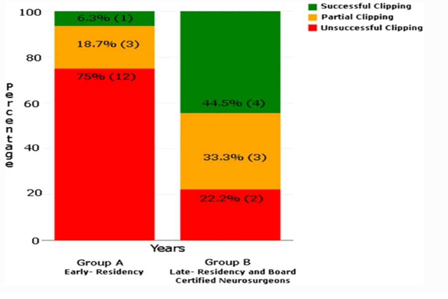
For the results of Group A and Group B, please refer to the chart above.
The nature of the editing (successful, unsuccessful, and partially completed) and the number of years of experience between the participants in groups A and B.
While 3D printing provides huge advantages to other industries such as aerospace, construction, and energy, it is particularly impressive in changing the face of medicine because it can provide more treatments for patients. Compared with traditional training types, this study provides good evidence. More than 80% of the participants thought the study was “a good choice for theoretical and traditional learning.”
“So far, neurosurgeons have only used one performance indicator: patient prognosis. The researchers concluded: “Our simulator can use other measures to improve and maintain skills. Further research is needed to confirm that the current simulator is a valuable clinical tool for measuring, evaluating and maintaining quality assurance in the training and education of the next generation of neurosurgeons. “
No one is willing to accept surgeons with little hands-on training for surgery, but throughout history, finding a suitable model or corpse has been a challenge for medical students. 3D printing has brought many benefits to doctors and patients around the world. 3D printed models and guides, whether showing organs such as kidneys, head and neck tumors or hip fractures, can be better diagnosed and treated, and provide comprehensive visual assistance for everyone involved.
Although the simulation equipment is more complicated, it is also very helpful for students and doctors. Now, Swiss medical researchers at the University of Bern have developed a new training method, detailed in the recently published “Neurosurgery Simulator for Training Aneurysm Microsurgery-For users involving neurosurgeons and residents Sex Research”. At present, due to the degree of difficulty involved in microsurgery, the training of intracranial aneurysms is limited. Medical students and surgeons usually gain experience through observation and assistance during the operation, performing operations under the supervision of skilled doctors, and learning videos. Although certain types of virtual reality simulations are available, researchers are still motivated to improve the technology using patient-specific models that even include functions such as blood circulation and pulsatility, which are very important for training purposes, but history It has been lacking.
Considering the subtle nature of intracranial aneurysm microsurgery, it is important for students to understand the vascular nature of the aneurysm and the exact condition in the event of a rupture. The training of students and doctors should have a comprehensive understanding of the surgical process and learn how to find blockages (blockages). Worryingly, due to the difficulty of model and other experiential education, there may continue to be a lack of well-trained cerebrovascular neurosurgeons.
The purpose of this research is to create a progressive simulator based on 3D printed models that will be able to train residents and neurosurgeons to trim intracranial aneurysms. The materials used are designed to mimic human tissue and include:
Unobstructed arterial wall
- .thickness
- .elasticity
- .Pulsating blood flow
Patient-specific 3D printed skull skull with patient brain model
Representation of the model in the training study: a patient-specific 3D printed skull skull with a model of the patient’s brain. b The MCA aneurysm model is located in the left fissure of Sylvian. c pulsate blood vessels and enter the pathological state of the model. d Brain retractor during operation by the resident.
25 neurosurgeons and residents participated in the study. Sixteen are in an early stage and have received less than 4 years of training in the field of neurosurgery; nine patients have been hospitalized late and have been certified by the board of directors to receive 4 to 15 years of neurosurgery training. Residents and surgeons use a five-point scale to score the following items to evaluate the effectiveness of the new simulator:
- .Surgery Simulated Anatomy
- .realism
- .Touch
- .Touch
- .General usage